
IGF-1 LR3 Research Peptide: Comprehensive Laboratory Guide
For professional laboratory research only. Not for human or veterinary use. Not for food, supplement, cosmetic, household, diagnostic, or therapeutic use.
Overview
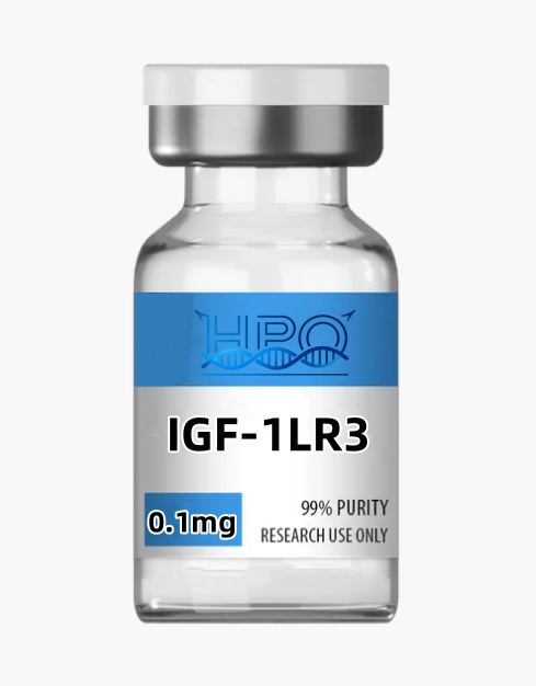
IGF-1 LR3 research peptide, also written Long R3 IGF-I, Long-[Arg3]IGF-I, or LR3IGF-I, is an engineered 83-amino-acid analogue of mature human insulin-like growth factor I (IGF-I). The literature-defined molecule contains the complete 70-residue human IGF-I chain with glutamate at native position 3 replaced by arginine, plus a 13-residue N-terminal extension. The modification was developed for biochemical systems in which native IGF-I activity is strongly shaped by insulin-like growth factor-binding proteins (IGFBPs). It is not endogenous IGF-I, ordinary recombinant human IGF-I, Des(1-3)IGF-I, R3-IGF-I without the extension, insulin, or an IGF-binding-protein complex.
The most defensible working model is that Long R3 retains activity at the type 1 IGF receptor (IGF1R) while binding much less strongly than native IGF-I to several IGFBPs. In an IGFBP-rich culture or biological matrix, reduced sequestration can increase the fraction of analogue available to interact with receptors. That does not mean the analogue has universally higher intrinsic affinity for IGF1R, a fixed biological half-life, or the same response in every cell type. Receptor abundance, IGFBP expression, insulin-receptor isoforms, hybrid receptors, ligand depletion, proteolysis, matrix adsorption, and exposure time all influence the observed signal.
Published Long R3 work is primarily nonclinical. Investigators have used the analogue to probe ligand availability, receptor signaling, cell-cycle responses, extracellular-matrix synthesis, intestinal physiology, lineage-specific differentiation, protein production, and bioanalytical detection. A recent mouse and cell study also tested Long R3 in an amyloid-model context and reported mixed outcomes rather than broad functional preservation. These studies define laboratory questions, not clinical benefits. This page therefore separates chemical identity, assay design, analytical quality, and evidence limits from unsupported treatment or performance claims.
Identity and physicochemical profile
| Catalog name | IGF-1 LR3 research peptide |
|---|---|
| Common research names | Long R3 IGF-I; Long-[Arg3]IGF-I; LR3IGF-I; IGF-1 LR3 |
| Molecular design | Human IGF-I analogue with a 13-residue N-terminal extension and an Arg substitution for Glu at native IGF-I position 3 |
| Chain length | 83 amino acids in the literature-defined mature analogue |
| N-terminal extension | MFPAMPLSSLFVN |
| Full literature sequence | MFPAMPLSSLFVNGPRTLCGAELVDALQFVCGDRGFYFNKPTGYGSSSRRAPQTGIVDECCFRSCDLRRLEMYCAPLKPAKSA |
| Approximate molecular mass | Approximately 9.1 kDa for the correctly oxidized, unmodified 83-residue protein; confirm the theoretical basis and observed intact mass in lot documentation |
| Database identifiers | CAS 143045-27-6; FDA GSRS UNII M9L22Y19H9. A registry identifier supports substance identification and does not imply regulatory approval. |
| Disulfide architecture | Three intramolecular disulfide bonds expected from the IGF-I fold; native IGF-I numbering is Cys6-Cys48, Cys18-Cys61, and Cys47-Cys52 |
| Protein class | Engineered, single-chain insulin-like growth factor analogue |
| Primary receptor context | IGF1R-centered research, with insulin-receptor isoforms and IGF1R/insulin-receptor hybrids considered where expressed |
| Binding-protein feature | Markedly reduced affinity for multiple IGFBPs compared with native IGF-I in published systems |
| Expression and processing | Source- and lot-dependent; host system, cleavage boundaries, terminal heterogeneity, folding, and purification must be documented |
| Purity and content | Lot-specific values only. Chromatographic area purity, intact-protein assay, total protein, water, counterions, aggregates, and active concentration are distinct measurements. |
| Intended use | Controlled laboratory research and analytical method development only; not for human or veterinary use |
The “Long” and “R3” elements describe two separate structural changes. R3-IGF-I contains the native-length chain with the Glu-to-Arg substitution; Long-IGF-I contains the extension without that substitution; Long R3 combines both. A primary rat study explicitly described R3-IGF-I as the position-3 substitution and Long-IGF-I as the 13-residue extension, while showing that the analogues remained active in an IGF receptor context despite reduced binding-protein interaction.[1] These forms must not be collapsed into a single identifier or used interchangeably as analytical standards.
Correct sequence alone is not sufficient to define an active IGF-like protein. The IGF-I fold contains three intramolecular disulfides, and recombinant production can generate alternative disulfide isomers that share the same amino-acid composition and nearly the same intact mass. Direct chemical-synthesis and recombinant-protein studies established the native IGF-I connectivity as Cys6-Cys48, Cys18-Cys61, and Cys47-Cys52.[2] In Long R3 numbering, the extension shifts those positions by 13 residues, but reporting the native numbering preserves comparability with established IGF-I literature.
The approximate mass is a screening attribute, not a complete identity test. Oxidation of six thiols into three disulfides changes the mass relative to the fully reduced chain, while methionine oxidation, deamidation, terminal clipping, adducts, retained leader sequence, and other process-related variants can alter the observed envelope. A qualified laboratory should therefore connect intact mass to sequence coverage, disulfide mapping, chromatographic behavior, aggregate assessment, and a content method.
IGF1R, IGFBP, and signaling framework
1. IGF1R binding and receptor autophosphorylation
IGF1R is a preformed receptor tyrosine kinase complex. Work with full-length and engineered receptors shows that ligand binding relieves an ectodomain constraint and permits transmembrane and intracellular rearrangements that support receptor autophosphorylation.[3] Earlier kinase-deficient receptor experiments demonstrated that IGF1R tyrosine-kinase activity is necessary for several IGF-I-initiated biochemical and cellular responses.[4] Long R3 is commonly used as an IGF1R-directed ligand in cell systems, but an observed downstream signal does not by itself prove exclusive action through IGF1R.
A robust receptor experiment should measure early receptor phosphorylation, total receptor abundance, downstream adapters, and a functional endpoint on separate time scales. Receptor knockdown or knockout, a validated blocking antibody, and a well-characterized kinase inhibitor can test pathway necessity, but each perturbation has limitations. A rescue experiment with restored IGF1R expression is stronger than inhibitor data alone. Ligand-depletion controls are useful when cell number or incubation time is high.
2. Reduced IGFBP association and matrix-dependent availability
The central experimental feature of Long R3 is reduced association with IGFBPs. Comparative studies reported roughly three orders of magnitude lower affinity than native IGF-I for several binding-protein preparations and linked this difference to greater apparent potency in IGFBP-producing cell systems.[5] Lung-fibroblast experiments similarly found a left-shifted proliferative response to Long R3 and showed that added IGFBP-3 inhibited IGF-driven cell-number changes.[6] These results support the availability model: lower IGFBP capture can leave more ligand accessible under defined conditions.
The magnitude of that effect is not portable across media. Serum lot, albumin, extracellular matrix, conditioned-medium volume, cell density, IGFBP proteases, and the expression of IGFBP-1 through -6 all matter. Serum-free conditions do not automatically remove binding proteins because cells may secrete them. Researchers should measure relevant IGFBPs, compare fresh and conditioned media, include native IGF-I, and test whether an added binding protein changes the rank order. “More potent” should always be attached to the exact assay rather than presented as an intrinsic universal property.
3. IRS, PI3K-AKT-mTOR, and RAS-MAPK pathway separation
After IGF1R phosphorylation, insulin-receptor substrates and SHC-family adapters can recruit PI3K-AKT and RAS-RAF-MEK-ERK modules. These branches overlap but are not interchangeable. In human chondrocytes, native IGF-I activated both AKT and ERK, yet inhibitor experiments associated proteoglycan synthesis with PI3K, mTOR, and p70S6K rather than a requirement for ERK.[7] In other cell types, ERK can contribute substantially to transcription or proliferation. The pathway supporting one endpoint cannot be assumed from a pathway diagram.
For Long R3 experiments, measure phospho-sites alongside total protein and loading controls, establish a short time course, and avoid treating one late phospho-blot as proof of sustained signaling. PI3K or MEK inhibitors require vehicle controls and independent confirmation of pathway suppression. IRS degradation and other feedback processes can change responsiveness during prolonged exposure. A receptor-proximal endpoint, a pathway-level endpoint, and a functional readout provide a more interpretable evidence chain.
4. Insulin receptor isoforms and hybrid-receptor confounding
Cells can express IGF1R, insulin receptor isoform A or B, and IGF1R/insulin-receptor hybrid complexes. Primary receptor studies show that ligand occupancy can drive phosphorylation across hybrid receptor subunits, complicating a simple one-ligand/one-receptor assignment.[8] Long R3 has also been used to study substrate activation in engineered insulin-receptor systems, reinforcing the need to characterize the receptor background rather than labeling every response “IGF1R specific.”[9]
Useful controls include receptor-null or single-receptor cell lines, quantified IGF1R and insulin-receptor expression, isoform-aware assays, and ligands with documented selectivity under the same conditions. Phospho-antibodies that recognize homologous IGF1R and insulin-receptor motifs may not distinguish them. Immunoprecipitation, targeted proteomics, or receptor-specific genetic perturbation can reduce ambiguity. Insulin contamination or endogenous IGF production should also be considered in complex media.
5. Cell proliferation, differentiation, and lineage dependence
Long R3 has been used in fibroblast, granulosa-cell, myoblast, and other culture systems to investigate proliferation or differentiation. Granulosa-cell work showed that Long R3 could stimulate IGFBP production and emphasized feedback between ligand action and binding-protein expression.[10] A cachexia-model study used Long R3 in myogenic experiments and reported differentiation-associated changes, but in the accompanying tumor-bearing mouse model the analogue also accelerated tumor growth.[11] That mixed result is an important evidence boundary, not a promotional endpoint.
Cell-number assays can reflect division, survival, altered metabolism, detachment, or assay interference. Use direct counts or DNA-based measurements with viability, apoptosis, cell-cycle, and morphology endpoints. For differentiation, include lineage markers, fusion or structural metrics, and normalization for baseline cell number. Transformed cell lines, primary cells, organoids, and tissues may respond differently because receptor abundance and feedback networks differ. Any proliferative signal requires containment and model-specific risk assessment.
6. Extracellular-matrix and tissue-physiology models
IGF-system ligands have been studied in cartilage and intestinal models. Work in osteoarthritic chondrocytes used R3-IGF-I as a reduced-IGFBP-binding comparator and found that disrupting IGF/IGFBP association could restore a matrix-synthesis response under the tested conditions.[12] A rat intestinal study reported that Long R3 changed some absorption measurements, but the authors attributed the finding mainly to altered mucosal mass rather than direct up-regulation of a specific transporter.[13] This distinction illustrates why morphology and tissue mass must be separated from per-cell function.
Matrix studies should quantify synthesis and degradation, normalize to viable cell or tissue content, and measure baseline matrix composition. Tissue-physiology studies need sham or vehicle controls, blinded histology, prespecified regions, and both absolute and normalized functional metrics. Results from native IGF-I cannot automatically be assigned to Long R3 because reduced IGFBP interaction changes distribution and exposure. Likewise, an increase in a tissue dimension is not equivalent to improved physiological function.
7. Neural, microglial, and amyloid-model research
A recent study tested Long R3 in male 5XFAD mice and in BV-2 microglial cells. The investigators reported changes in aspects of cortical amyloid-plaque composition and microglial uptake-associated pathways, while multiple behavioral and memory measures were not preserved.[14] The value of this study is its mixed outcome: a molecular or histological shift did not translate into a broad functional conclusion. It does not establish prevention or treatment of neurodegenerative disease.
Replication would require sex-inclusive designs, blinded pathology, well-characterized amyloid species, microglial state markers, exposure verification, and controls for metabolic or systemic changes. BV-2 cells are not equivalent to primary microglia or an intact brain. Plaque morphology, soluble oligomers, inflammatory transcription, synaptic endpoints, and behavior should remain separate outcomes. The analogue’s effect on one compartment cannot be generalized to cognition or clinical status.
Appropriate nonclinical research applications
Within qualified facilities, the IGF-1 LR3 research peptide may support narrowly framed laboratory programs such as:
- Ligand-availability experiments: compare Long R3 with native IGF-I in defined media containing endogenous, added, or depleted IGFBPs.
- Receptor-proximal signaling: measure IGF1R autophosphorylation, adapter recruitment, and early pathway kinetics in receptor-characterized systems.
- Pathway-dissection studies: test PI3K-AKT-mTOR and RAS-MAPK requirements with genetic and pharmacological perturbations.
- Cell-state research: examine proliferation, survival, cell cycle, differentiation, or matrix production with orthogonal endpoints and lineage controls.
- IGFBP feedback studies: quantify how ligand exposure changes IGFBP transcription, secretion, proteolysis, and subsequent ligand availability.
- Receptor-selectivity models: distinguish IGF1R, insulin-receptor isoforms, and hybrid-receptor contributions using engineered cells and receptor-specific measurements.
- Protein analytical development: establish intact-mass, peptide-map, disulfide-map, chromatographic, aggregate, and activity methods for the 83-residue analogue.
- Recombinant-production research: evaluate expression host, cleavage, folding, disulfide formation, purification, and lot-to-lot comparability.
These are experimental categories, not validated protocols or indications. Related catalog materials can help define orthogonal controls: AICAR metabolism research, NAD+ analytical research, MOTS-c signaling research, Humanin peptide research, and SS-31 mitochondrial research involve distinct molecular systems. Internal links support navigation only and do not imply compatibility, co-use, or synergy.
Experimental design and controls
- Define the molecular article. Record the full sequence, terminal state, expression host, processing boundary, disulfide assignment, lot, CoA version, and content basis. Do not infer Long R3 identity from the product name alone.
- Characterize the receptor background. Quantify IGF1R, insulin-receptor isoforms, and relevant hybrid receptors where feasible. Confirm that detection antibodies distinguish intended targets.
- Map the IGFBP environment. Document serum source, conditioned-medium history, IGFBP expression, albumin, and extracellular-matrix components. Include native IGF-I and defined IGFBP controls when testing the Long R3 design hypothesis.
- Use a concentration-time matrix. Select in vitro ranges from assay performance, ligand recovery, receptor occupancy questions, and model tolerance. Avoid extrapolating from nominal concentration without exposure measurement.
- Build receptor causality. Combine receptor-specific genetic perturbation with a pharmacological or antibody control. Confirm pathway engagement upstream and downstream of the perturbation.
- Separate response classes. Measure proliferation, survival, metabolism, differentiation, matrix synthesis, and morphology independently. One colorimetric signal cannot stand for all of them.
- Control handling artifacts. Evaluate adsorption to tubes and plates, proteolysis, oxidation, aggregation, freeze-thaw effects, filtration recovery, and carryover in the actual matrix.
- Use biological and technical replicates correctly. Wells from one preparation are technical replicates, not independent biological samples. Predefine exclusions, normalization, primary endpoints, and multiplicity correction.
- Report negative and mixed findings. A molecular change without a functional effect is informative. Avoid selecting only growth-associated endpoints or presenting nonclinical associations as clinical evidence.
For broader peptide-system comparisons, laboratories may review Ipamorelin research, Sermorelin acetate research, Tesamorelin research, PEG-MGF research, Cerebrolysin research, and Epithalon peptide research. None is an interchangeable IGF1R control. Comparative work requires identity-confirmed single-material arms and a prespecified mechanistic reason for inclusion.
Analytical identity and quality control
Quality assessment must separate identity, purity, quantity, conformation, aggregation, and biological activity. A single reverse-phase HPLC area percentage cannot establish all six. For the IGF-1 LR3 research peptide, a fit-for-purpose analytical package may include:
- Intact-protein mass: qualified LC-MS or high-resolution MS matched to the declared 83-residue oxidized chain, with deconvolution settings, mass tolerance, adduct handling, and raw spectra.
- Sequence confirmation: reduced and alkylated peptide mapping with adequate coverage of the N-terminal extension, the Arg3 substitution site, termini, and process-sensitive regions.
- Disulfide mapping: nonreduced peptide mapping or another orthogonal method that distinguishes native connectivity from alternative isomers. Intact mass alone cannot establish bond pairing.
- Chromatographic purity: a stability-indicating reversed-phase method with system suitability, integration rules, reporting threshold, and evidence that oxidized, clipped, and misfolded species are resolved where relevant.
- Aggregation and size variants: size-exclusion chromatography, field-flow fractionation, analytical ultracentrifugation, light scattering, or another suitable method, recognizing that dilution and column interaction can change apparent aggregates.
- Protein content: calibrated amino-acid analysis, isotope-dilution mass spectrometry, nitrogen analysis, or a qualified spectroscopic method. Report content separately from chromatographic area purity and gross vial mass.
- Higher-order structure: circular dichroism, spectroscopy, thermal methods, or receptor-binding comparison when the study requires conformational comparability.
- Biological activity: a receptor-proximal cell assay with a traceable comparator, defined receptor background, linear response range, and acceptance criteria. Bioactivity does not replace chemical identity.
- Model-specific attributes: endotoxin, bioburden, host-cell proteins, residual DNA, process reagents, or compatibility testing when relevant to the planned laboratory system. Such testing does not make material suitable for administration.
Recombinant Long R3 production has been demonstrated in engineered expression systems, including recent work using a fusion strategy followed by purification and cell-based activity assessment.[15] That literature establishes production feasibility, not equivalence among commercial lots. Host-cell background, protease cleavage, N-terminal heterogeneity, oxidation, refolding conditions, and purification determine the impurity profile. A reference standard should have its own sequence, disulfide, water, content, and traceability documentation.
Mass-spectrometric antidoping research has distinguished Long R3, R3-IGF-I, Des(1-3)IGF-I, and N-terminal Long R3 degradation products by immunopurification and high-resolution MS.[16] This illustrates two practical points: closely related IGF analogues need sequence-specific detection, and clipped forms may persist differently from intact protein. A method that detects a shared internal peptide without an extension-specific peptide may misassign identity.
Stability-indicating development should investigate plausible pathways rather than declare a generic shelf life. Relevant targets include methionine oxidation, asparagine or glutamine deamidation, peptide-bond clipping, disulfide scrambling, dimerization, higher-order aggregation, surface adsorption, and loss of receptor activity. Forced-condition studies should be proportionate and used to demonstrate method specificity, not to predict routine stability directly. Lot release and ongoing suitability must be based on the matching CoA and actual storage history.
Handling, storage, and documentation
Handle the IGF-1 LR3 research peptide only in qualified laboratories using trained personnel, suitable personal protective equipment, engineering controls, and an approved chemical-hygiene or biosafety plan. Review the current safety data sheet and lot certificate before opening. Prevent inhalation of dry material, unintended contact, cross-contamination, and environmental release. Keep research material segregated from medicines, food, cosmetics, personal items, and clinical supplies.
Storage requirements are lot- and presentation-specific. Follow the condition printed on the matching label and CoA rather than a generic web statement. Laboratories should qualify the actual container, temperature, humidity, light exposure, headspace, matrix, concentration, and handling cycle. Low protein concentration can increase surface loss, while repeated thermal or mechanical stress can change aggregation or folding. Compatibility with glass, polymer, filters, tubing, and assay plates must be measured rather than assumed.
This page intentionally provides no dosing, injection, administration, or reconstitution procedure. Analytical or cell-culture preparation should follow an institution-approved SOP that documents solvent or matrix identity, pH, ionic strength, mixing history, hold time, container, concentration basis, and recovery. Nominal mass is not automatically active protein content. Calculations should use lot-specific protein assay and, where relevant, correct for water, counterions, excipients, and nonprotein material.
Retain the receipt record, chain of custody, lot CoA, raw chromatograms, mass spectra, peptide maps, disulfide data, aggregate results, preparation worksheet, instrument sequence, storage log, and deviations. Report the exact lot and molecular form in publications. The site’s research-use terms describe the general intended-use boundary, but only matching lot documents can support a batch-specific claim.
Evidence and regulatory boundary
Long R3 evidence consists mainly of cell, tissue, analytical, and animal studies. Many experiments use the analogue as a tool to reduce IGFBP confounding rather than as a candidate medicine. Even within nonclinical work, outcomes vary by species and model. For example, a pig study reported reductions rather than growth promotion in several physiological measures, despite the analogue’s greater apparent activity in other systems.[17] This variability directly contradicts universal anabolic or performance claims.
FDA-approved mecasermin is a different molecular article. The current U.S. label describes Increlex as recombinant human IGF-I with a 70-amino-acid sequence identical to endogenous human IGF-I and three intramolecular disulfide bridges.[18] Long R3 contains 83 residues and a sequence substitution. Approval, clinical labeling, manufacturing controls, dosage information, or safety conclusions for mecasermin do not transfer to this catalog analogue. This product is not represented as an FDA-approved drug, biosimilar, interchangeable biologic, food, supplement, or cosmetic.
FDA’s Global Substance Registration System lists Long-(Arg3)insulin-like growth factor-I under UNII M9L22Y19H9 and maps CAS 143045-27-6, while expressly separating substance registration from regulatory approval.[19] For sport-governance context, the 2026 World Anti-Doping Agency Prohibited List places IGF-1 and its analogues in section S2.3 and prohibits them at all times, both in and out of competition.[20] These identity and antidoping records do not establish human safety or efficacy.
IGF-axis biology also creates safety-relevant laboratory boundaries. Signaling can affect glucose handling, cell survival, proliferation, and tumor-model behavior. The cachexia study’s tumor-growth result and the amyloid study’s failure to preserve behavioral outcomes show why pathway engagement cannot be marketed as a human benefit. Human self-experimentation, athletic or bodybuilding use, growth promotion, anti-aging use, diagnosis, treatment, or prevention are outside the intended use.
A CAS number, approximate mass, sequence string, or generic “research grade” label does not establish lot identity, sterility, clinical safety, receptor selectivity, or legal status for administration. Evidence for native IGF-I or an approved rhIGF-I product cannot be silently substituted for Long R3 data. Claims should identify the exact ligand, model, matrix, endpoint, and evidence level.
Frequently asked questions
What is the IGF-1 LR3 research peptide?
It is a literature-defined 83-amino-acid analogue of mature human IGF-I. It combines a 13-residue N-terminal extension with an Arg-for-Glu substitution at native IGF-I position 3. The analogue is used mainly to study IGF1R-related signaling and the effect of reduced IGFBP binding in controlled laboratory models.
Why is it called Long R3?
“Long” refers to the N-terminal extension, while “R3” refers to arginine replacing glutamate at position 3 of the native IGF-I portion. Long R3, R3-IGF-I, Long-IGF-I, Des(1-3)IGF-I, and native IGF-I are distinct analytical identities and should not share an unqualified sequence, standard, or result.
Does reduced IGFBP binding mean higher IGF1R affinity?
No. Published work supports markedly reduced association with multiple IGFBPs and increased apparent activity in some IGFBP-rich systems.[5] That effect reflects ligand availability and does not prove universally higher intrinsic receptor affinity. The outcome depends on the receptor and binding-protein environment.
How should receptor specificity be tested?
Characterize IGF1R, insulin-receptor isoforms, and hybrid receptors; combine receptor-specific genetic perturbation with an orthogonal blocker; and measure receptor-proximal phosphorylation plus downstream and functional endpoints. A phospho-AKT or proliferation signal alone cannot identify the initiating receptor.
Why are disulfide mapping and aggregation tests important?
The IGF-I fold requires three specific intramolecular disulfides. Alternative pairings can preserve amino-acid composition and similar intact mass while changing conformation or activity. Aggregates and oligomers are also not captured reliably by a single reversed-phase purity result. Nonreduced mapping and a size-variant method address different quality questions.
What purity, content, or shelf life does this page guarantee?
No generic value is asserted. Area purity, protein assay, aggregate level, water, gross fill mass, active concentration, storage period, and availability are lot-specific. Review the current variant and matching CoA. A high HPLC area percentage is not equivalent to correct folding, net content, sterility, or biological activity.
Is IGF-1 LR3 intended for treatment, growth promotion, or performance use?
No. The catalog material is for controlled laboratory research and analytical use only. It is not for human or veterinary use, injection, self-experimentation, bodybuilding, growth promotion, anti-aging, food, supplements, diagnosis, treatment, or disease prevention.
References
- Prosser CG, et al. Several IGF-I analogues and IGF/IGFBP-3 complexes fail to mimic growth hormone effects in a rat lactation model. J Endocrinol. 1994;140(2):211-219. Defines R3 and Long modifications and reduced IGFBP binding. PMID: 7513341.
- Iwai M, et al. Direct identification of disulfide bond linkages in human IGF-I by chemical synthesis. J Biochem. 1989;106(6):949-951. Establishes native IGF-I disulfide connectivity.
- Kavran JM, et al. How IGF-1 activates its receptor. eLife. 2014;3:e03772. Ligand-dependent IGF1R conformational and autophosphorylation mechanism. PMID: 25255214.
- Kato H, et al. Role of tyrosine kinase activity in signal transduction by the IGF-I receptor. J Biol Chem. 1993;268(4):2655-2661. Kinase-deficient receptor study. PMID: 7679099.
- Tomas FM, et al. Effects of interactions between IGFBPs and IGFs on plasma clearance and biological activities of IGF analogs. Endocrinology. 1993. Comparative IGF-I and LR3IGF-I binding and activity. PMID: 7683526.
- Goldstein RH, et al. IGFBP expression alters IGF-mediated proliferation of postnatal lung fibroblasts. Exp Lung Res. 2004. Long R3 and IGFBP-3 cell-system comparison. PMID: 15204833.
- Starkman BG, et al. IGF-I stimulation of proteoglycan synthesis by chondrocytes requires PI3K but not ERK MAPK. Biochem J. 2005;389:723-729. Pathway-separation experiment. PMID: 15801908.
- Frattali AL, Pessin JE. Ligand occupancy and autophosphorylation in insulin/IGF-1 hybrid receptors. J Biol Chem. 1993;268:7393-7400. Hybrid-receptor signaling context. PMID: 8463272.
- Denley A, et al. Differential activation of IRS-1 and IRS-2 by IGF-activated insulin receptors. Mol Cell Biol. 2007;27(9):3569-3577. Long R3 in receptor-engineered systems. PMID: 17325037.
- Adashi EY, et al. Insulin and IGFs stimulate IGF-binding-protein production by ovarian granulosa cells. Endocrinology. 1992;131(1):329-336. Ligand/IGFBP feedback and Long R3 comparison. PMID: 1379161.
- Hatakeyama S, et al. Inhibition of activin-like kinase 4/5 attenuates cancer-cachexia-associated muscle wasting. Sci Rep. 2019;9:11384. Long R3 myogenic and tumor-model findings. PMID: 31285507.
- De Ceuninck F, et al. Disruption of IGF-1 binding to IGFBPs restores responses in osteoarthritic chondrocytes. Arthritis Res Ther. 2004;6:R393-R403. R3-IGF-I as a reduced-binding-protein comparator.
- Garnaut SM, et al. Effects of IGF-I and Long-R3-IGF-I on intestinal glucose-analogue absorption and mucosal responses. Growth Factors. 2002;20(1):17-25. Tissue mass versus transport interpretation. PMID: 11999215.
- Intranasal Long R3 IGF-1 promotes amyloid-plaque remodeling but fails to preserve cognitive function in male 5XFAD mice. J Alzheimers Dis. 2024. Mixed neural-model outcomes. PMID: 39610283.
- Recombinant expression of IGF-1 and LR3 IGF-1 fused with xylanase in Pichia pastoris. Appl Microbiol Biotechnol. 2023. Recombinant-production and bioactivity study. PMID: 37261455.
- Detection of LongR3-IGF-I, Des(1-3)-IGF-I, and R3-IGF-I using immunopurification and high-resolution mass spectrometry. Drug Test Anal. 2021. Analogue-specific detection and degradation products. PMID: 33587816.
- Dunaiski V, et al. Long [R3] IGF-I reduces growth and several endocrine measures in pigs. J Endocrinol. 1997;155(3):559-565. Species- and model-dependent outcome. PMID: 9488001.
- U.S. National Library of Medicine. DailyMed: Increlex (mecasermin) current prescribing information. Defines the approved 70-residue recombinant human IGF-I product, which is chemically distinct from Long R3.
- U.S. Food and Drug Administration. GSRS substance record for Long-(Arg3)insulin-like growth factor-I. Provides UNII M9L22Y19H9 and CAS 143045-27-6; UNII assignment does not imply approval.
- World Anti-Doping Agency. The 2026 Prohibited List, S2.3. Lists IGF-1 and its analogues as prohibited at all times.
Research-use-only notice: This page summarizes chemical identity, analytical considerations, and published nonclinical research for qualified laboratory planning. It is not medical advice and provides no administration instructions. The product is not intended for human consumption or veterinary use, injection, diagnosis, treatment, prevention, bodybuilding, growth promotion, anti-aging, or performance enhancement.


Reviews
There are no reviews yet.