
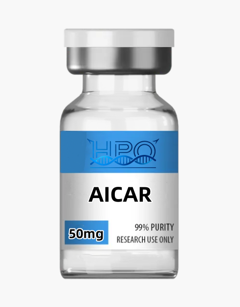
AICAR Research Compound: Comprehensive Laboratory Guide
For professional laboratory research only. Not for human or veterinary use. Not for food, supplement, cosmetic, household, diagnostic, or therapeutic use.
Overview
AICAR research compound is the small-molecule nucleoside 5-aminoimidazole-4-carboxamide riboside, also known as AICA riboside, AICAr, acadesine, or Z-riboside. It is widely used as an experimental probe of cellular energy sensing because many cells transport the riboside and phosphorylate it to 5-aminoimidazole-4-carboxamide ribonucleotide, commonly called ZMP. ZMP can reproduce several regulatory effects of AMP, including activation of AMP-activated protein kinase (AMPK).[3]
Despite its frequent appearance in peptide catalogs, acadesine is not a peptide. It is a ribonucleoside composed of an aminoimidazole carboxamide base linked to a ribofuranose. The abbreviation also requires care: many experimental papers call the extracellular riboside “AICAR,” while strict biochemical nomenclature reserves AICAR for the phosphorylated ribonucleotide that is an intermediate in de novo purine biosynthesis. This guide uses “AICAR research compound” for the commercial acadesine/AICA-riboside material and “ZMP” for its intracellular monophosphate unless discussing a paper’s original terminology.
AICAR is often described as an “AMPK activator,” but that label is incomplete. Cellular response depends on nucleoside transport, adenosine-kinase activity, ZMP accumulation, ATP and phosphate status, cell lineage, medium composition, exposure time, and metabolism of ZMP toward inosine monophosphate. ZMP also influences AMP-sensitive enzymes and nucleotide pathways independently of AMPK. Genetic loss-of-function studies have shown that some phenotypes persist without the relevant AMPK catalytic subunits.[6][7] Accordingly, AICAR is best treated as a metabolism-active nucleoside probe, not as a selective molecular switch or a substitute for genetic validation.
Molecular identity and physicochemical profile
| Catalog name | AICAR research compound |
|---|---|
| Preferred chemical identity | Acadesine; 5-aminoimidazole-4-carboxamide riboside |
| Common synonyms | AICA riboside; AICAr; Z-riboside; 5-amino-1-β-D-ribofuranosylimidazole-4-carboxamide |
| Chemical class | Purine-pathway nucleoside analogue; aminoimidazole ribonucleoside; not a peptide |
| Molecular formula | C9H14N4O5 for the neutral, unsolvated parent |
| Average molecular mass | 258.234 g/mol |
| Monoisotopic mass | 258.09642 Da |
| Expected positive ion | [M+H]+ calculated m/z approximately 259.1037 for the parent molecule; observed ions and adducts depend on method and matrix |
| CAS Registry Number | 2627-69-2 |
| Database identifiers | PubChem CID 17513; ChEBI CHEBI:28498; FDA UNII 53IEF47846 |
| InChIKey | RTRQQBHATOEIAF-UUOKFMHZSA-N |
| Identity boundary | ZMP/AICA ribonucleotide is a different, phosphorylated chemical entity: CAS 3031-94-5, C9H15N4O8P, average mass 338.213 g/mol, ChEBI CHEBI:18406. It is not the CAS 2627-69-2 catalog parent. |
| Stereochemical form | β-D-ribofuranosyl configuration as defined for acadesine |
| Physical form | Lot dependent; consult the certificate of analysis (CoA) rather than inferring appearance, hydration, or fill composition from a generic listing |
| Purity and content | Lot-specific values only. Chromatographic area purity, chemical identity, and net content are separate attributes and require separate evidence. |
| Catalog format | Confirm the currently selected product configuration, vial count, and labeled fill against the live variant and lot documents before ordering or entering the material into an inventory system. |
| Intended use | Analytical, biochemical, and in vitro laboratory research only; no use in humans or animals |
PubChem and ChEBI list the parent acadesine structure, formula, mass, identifiers, and stereochemistry; ChEBI separately records the phosphorylated AICA ribonucleotide.[1] Those values do not describe every commercial form. A hydrate, solvate, salt, isotopically labeled analogue, or mixture with excipients will have a different complete mass balance. Researchers should distinguish the 258.234 g/mol riboside from 338.213 g/mol ZMP. A mass spectrum matching the riboside cannot demonstrate that a biological sample accumulated ZMP; intracellular nucleotide analysis must measure that metabolite directly.
Ribose hydroxyls make acadesine polar, while the aminoimidazole carboxamide system provides UV absorbance and hydrogen-bonding sites. Apparent solubility, retention, ionization efficiency, and degradation rate vary with pH, ionic strength, solvent composition, temperature, and concentration. LC–MS methods should assess matrix suppression, sodium or potassium adducts, in-source fragmentation, and separation from structurally related nucleosides.
Mechanistic and research themes
1. Nucleoside transport and intracellular phosphorylation
The extracellular riboside must generally enter a cell before the canonical ZMP mechanism can occur. Work in primary mouse hepatocytes found that inhibiting equilibrative nucleoside transporter 1 (ENT1) impeded acadesine disappearance from the medium and blocked its effects on hepatic glucose production and AMPK-associated readouts.[4] After transport, adenosine kinase phosphorylates the riboside to ZMP. The importance of this conversion can be tested with transporter inhibitors, adenosine-kinase perturbation, or direct LC–MS measurement of intracellular acadesine (AICAr), ZMP, AMP, ADP, and ATP.
This step creates a major source of model dependence. Two cell lines exposed to the same nominal extracellular concentration can accumulate very different amounts of ZMP because they differ in transporter expression, kinase activity, cell density, nucleotide turnover, or medium nucleosides. Failure to see AMPK phosphorylation does not necessarily mean the supplied compound lacks identity, and a strong response does not prove direct binding of the riboside to AMPK. Exposure should be established, not assumed.
2. AMP mimicry and AMPK pathway engagement
The landmark intact-cell study by Corton and colleagues showed that ZMP accumulated in rat hepatocytes and reproduced both allosteric and phosphorylation-dependent activation of AMPK, with downstream inhibition of fatty-acid and sterol synthesis under those conditions.[3] In current assay language, useful proximal readouts include AMPK Thr172 phosphorylation and activity plus phosphorylation of a validated substrate such as acetyl-CoA carboxylase (ACC). Neither readout is sufficient by itself: phosphorylation can diverge from activity, antibodies require specificity controls, and ACC can be influenced by additional signals.
To assign a phenotype to AMPK, combine AICAR with genetic deletion or knockdown of the relevant AMPK catalytic subunit, rescue with a defined construct, and a chemically distinct AMPK modulator. Compound C/dorsomorphin alone is not a definitive AMPK control because it has multiple kinase and cellular effects. The strongest design establishes intracellular ZMP, confirms proximal pathway engagement, and then asks whether the downstream phenotype disappears when AMPK signaling is specifically disabled.
3. ACC, malonyl-CoA, and lipid-flux experiments
AMPK-mediated phosphorylation of ACC can lower malonyl-CoA formation and alter the constraint on mitochondrial fatty-acid entry. Early hepatocyte studies also reported suppression of fatty-acid and sterol synthesis after acadesine exposure.[3] This makes the compound useful in tracer studies examining lipid synthesis, oxidation, and substrate choice. However, effects are not universal across tissues, nutritional states, or exposure conditions, as summarized in the systematic review.[2]
Researchers should distinguish enzyme phosphorylation from net pathway flux. Recommended endpoints include isotopic substrate tracing, acylcarnitine profiles, oxygen-consumption measurements, malonyl-CoA quantitation, and direct measurement of cellular energy charge. A decrease in a lipid pool can reflect altered synthesis, oxidation, export, uptake, or cell number. Normalization to viable cell mass and time-resolved sampling reduce interpretive ambiguity.
4. Glucose transport and transporter trafficking
AICAR has been used extensively in skeletal-muscle systems to examine insulin-independent glucose transport. In a rat muscle study, the compound increased glucose uptake, recruited GLUT4 to the plasma membrane but not the transverse-tubule fraction, and activated p38 MAPK; inhibition of p38 reduced the uptake response.[5] Other work implicates AMPK-dependent phosphorylation of trafficking regulators such as TBC1D1, yet the size and direction of the response vary with fiber type, species, culture state, and assay design.
These data do not justify describing AICAR as a general glucose-lowering agent. Isolated cardiomyocytes, for example, have shown AMPK activation without the expected GLUT4-mediated uptake response. A rigorous experiment separates transporter abundance, surface localization, substrate uptake, glycolytic flux, and insulin signaling rather than treating them as interchangeable. Use a transportable glucose analogue with appropriate controls, verify cell viability, and measure the relevant transporter compartment directly.
5. mTOR, protein synthesis, and autophagy context
Because AMPK can oppose anabolic signaling through TSC2, raptor, and related nodes, acadesine is frequently used to test AMPK–mTOR pathway relationships. Published studies report context-dependent changes in protein synthesis and mTOR-associated signaling.[2] In cell culture, phosphorylation of S6K, S6, or 4E-BP1 can be paired with puromycin-incorporation or isotope-based protein-synthesis assays. Autophagy claims require flux measurements—such as LC3 turnover with and without lysosomal blockade—rather than a static LC3 or p62 value.
Interpretation must remain context specific. Altered nucleotide pools, ATP depletion, stress responses, and cell-cycle changes can affect translation independently of canonical AMPK signaling. AICAR may produce different autophagy phenotypes depending on tissue, exposure duration, nutrient status, and disease model. It should not be described simply as an autophagy “inducer” or “inhibitor” without a validated flux experiment and pathway-specific controls.
6. PGC-1α, mitochondrial programs, and exercise-model literature
Longer acadesine exposures have been used to study transcriptional programs related to oxidative metabolism, PGC-1α, and mitochondrial biogenesis. A 2008 mouse study reported that repeated exposure increased selected oxidative genes and endurance in sedentary mice, helping popularize the “exercise mimetic” label.[8] This is a preclinical observation, not evidence of safety, effectiveness, or an appropriate performance use in humans.
Those studies are preclinical and do not make AICAR equivalent to exercise. Exercise integrates mechanical loading, neural input, endocrine signals, perfusion, temperature, and tissue cross-talk that a single compound does not reproduce. In vitro mitochondrial-biogenesis studies should combine mitochondrial DNA copy number, organelle imaging, respiratory capacity, protein abundance, and citrate-synthase activity. An increase in one transcript is not sufficient evidence of more functional mitochondria, and an increase in mitochondrial mass does not guarantee improved coupling or ATP production.
7. Purine biosynthesis and nucleotide-pool remodeling
ZMP is not merely an AMPK ligand mimic; it is also a native intermediate in de novo purine synthesis. The bifunctional enzyme ATIC normally converts ZMP toward formyl-AICAR and inosine monophosphate. Exogenous AICA riboside can therefore interact with purine-pathway capacity, one-carbon metabolism, adenosine handling, and the abundance of adenine nucleotides. Depending on cell type and time, the compound can change ATP, AMP, inorganic phosphate, or other metabolites that independently influence signaling.
This dual identity is especially important in proliferation and stress studies. A change in cell growth may reflect AMPK, nucleotide imbalance, bioenergetic stress, or several of these processes together. Targeted metabolomics should include ZMP and adenine nucleotides, and ideally IMP, inosine, adenosine, and pathway-relevant intermediates. ATIC status, folate availability, and cell-cycle distribution are informative covariates. The 2021 systematic review catalogues many AMPK-independent effects and is a useful guide to potential confounders.[2]
8. AMPK-independent actions and direct metabolic interference
Multiple experiments show why AICAR cannot serve as sole proof of AMPK causality. In liver-specific AMPK-knockout mice, acute suppression of glucose production persisted despite removal of hepatic AMPK, while energy stress was more pronounced.[6] In isolated hepatocytes and mitochondria, ZMP accumulation and phosphate depletion were associated with AMPK-independent inhibition of oxidative phosphorylation, including an effect on respiratory-chain complex I.[7] These findings demonstrate that a phenotype can reflect several routes even when AMPK phosphorylation rises in parallel.
These findings do not mean AMPK is irrelevant. They mean that the causal route must be established in each model. Include AMPK-null or knockdown cells where feasible, compare a structurally unrelated direct AMPK activator, quantify energy charge, and test whether adenosine-kinase inhibition changes both ZMP formation and phenotype. When respiration changes, examine substrate-specific complex activity, membrane potential, cell integrity, and nucleotide depletion before assigning the result to mitochondrial biogenesis or damage.
Appropriate nonclinical research applications
Within a qualified laboratory and after a task-specific chemical risk assessment, AICAR research compound can support the following analytical and in vitro programs:
- AMPK pathway assay development: use as a positive or challenge control for AMPK phosphorylation, kinase activity, ACC phosphorylation, and downstream transcription, paired with genetic and orthogonal chemical controls.
- Nucleoside transport and phosphorylation studies: quantify extracellular disappearance, ENT-family dependence, adenosine-kinase dependence, and intracellular ZMP formation using validated LC–MS methods.
- Metabolic-flux research: examine glucose uptake, glycolytic output, lipid synthesis, fatty-acid oxidation, and substrate preference with isotope tracers rather than endpoint abundance alone.
- Mitochondrial adaptation and stress assays: evaluate respiratory capacity, coupling, mitochondrial mass, PGC-1α-associated expression, and the possibility of direct respiratory inhibition across time and concentration.
- Purine and one-carbon pathway research: investigate ZMP handling, ATIC-dependent metabolism, adenine-nucleotide pools, and interactions between nutrient status and de novo purine synthesis.
- Cell-growth and translation models: test AMPK–mTOR hypotheses, protein-synthesis rates, cell-cycle distribution, and viability while separating AMPK-dependent signaling from nucleotide or energy stress.
- Analytical-method and stability studies: develop HPLC, LC–UV, LC–MS/MS, NMR, degradation, adsorption, freeze–thaw, and matrix-recovery methods for acadesine and ZMP.
These are research themes, not validated protocols or claims of efficacy. This catalog material is not intended for athletic enhancement, self-experimentation, clinical investigation, or administration to any person or animal. Historical animal or clinical literature cited for scientific context does not change the intended use of the supplied material.
Clinical, regulatory, and sports evidence boundary
Acadesine has a historical clinical-development record, but it did not demonstrate benefit in the large RED-CABG randomized trial. A prespecified futility analysis stopped enrollment after 3,080 participants had been randomized; the primary composite outcome occurred in 5.0% of the placebo group and 5.1% of the acadesine group, with no improvement in key secondary endpoints.[9] This negative result is important context and precludes any claim of established cardioprotection.
FDA records show an orphan-drug designation for acadesine, but FDA orphan-drug designation is not marketing approval. The FDA orphan record lists acadesine as not FDA approved for that orphan indication, and an FDA UNII is a substance identifier rather than evidence of regulatory review, safety, efficacy, or product approval.[10] This catalog research reagent is not an FDA-approved drug product.
The World Anti-Doping Agency’s 2026 Prohibited List expressly names AICAR in section S4.4.1 among AMPK activators. It is prohibited at all times, meaning both in-competition and out-of-competition.[11] The material must not be marketed, supplied, or used for athletic performance enhancement.
Analytical quality control
For a small polar nucleoside, release testing should separate four questions: Is the structure correct? What related substances are present? How much parent compound is actually in the container? Does the material perform consistently in the chosen analytical system? A single HPLC area percentage cannot answer all four. A fit-for-purpose quality package may include:
- Identity: high-resolution LC–MS matched to the declared parent or salt form, with accurate mass, isotope pattern, adduct assignments, instrument tolerance, and traceable raw-data identifiers.
- Orthogonal structure confirmation: qualified 1H NMR and, when needed, 13C NMR, two-dimensional NMR, optical rotation, or comparison with a traceable reference standard to address connectivity and ribose stereochemistry.
- Chromatographic purity: a stability-indicating HPLC or UPLC method with adequate retention for a polar nucleoside, stated column chemistry, mobile phases, detection wavelength, integration rules, reporting threshold, and system-suitability criteria.
- Net content: a calibrated quantitative method such as qNMR, mass-balance assay, or reference-standard HPLC. Report net acadesine content separately from area purity and gross fill mass.
- Water, solvent, and inorganic residues: Karl Fischer water where appropriate, residual-solvent testing based on process knowledge, and counterion or elemental testing when relevant to synthesis and isolation.
- Biological-assay compatibility: endotoxin, bioburden, or mycoplasma testing only when the intended in vitro system requires it. These tests do not establish sterility or suitability for administration.
Method suitability should be demonstrated across the intended range and matrix. Specificity is particularly important because acadesine, adenosine, inosine, and nucleotide metabolites may coexist in cell extracts. Stable-isotope internal standards, matrix-matched calibration, recovery experiments, and evaluation of ion suppression strengthen quantitative LC–MS work. The FDA’s final ICH Q2(R2) guidance provides a recognized framework for analytical-procedure validation, including specificity, accuracy, precision, range, and robustness; applying that framework voluntarily does not make a research reagent a pharmaceutical product.[12]
Experimental design and interpretation
- Confirm the test article. Record catalog and lot numbers, chemical form, purity method, net content, water, storage history, and opening date. Verify identity before interpreting a biological response.
- Measure intracellular exposure. Nominal medium concentration is not the same as intracellular acadesine (AICAr) or ZMP. Establish uptake and ZMP accumulation over the actual experimental interval.
- Use a concentration–time matrix. Select an in vitro range from solubility, recovery, viability, and pilot pathway data. Avoid importing a concentration from a different cell line without checking transport and metabolism.
- Predefine the causal question. Separate AMPK engagement, metabolic flux, mitochondrial function, cell growth, and transcription as distinct outcomes. Choose one primary endpoint and a statistical plan before collecting data.
- Build orthogonal controls. Include vehicle, untreated, assay-positive, structurally unrelated AMPK activator, transporter or kinase perturbation, AMPK loss-of-function, and rescue controls where technically appropriate.
- Track energy and nucleotide state. Measure ATP, ADP, AMP, ZMP, and ideally phosphate or related purine intermediates. These data often explain apparent off-target or stress phenotypes.
- Control assay interference. Run cell-free compound-plus-reagent wells for fluorescence, luminescence, colorimetric, and redox assays. Confirm major findings using a chemically distinct readout.
- Design for replication. Use independent biological replicates, randomized plate positions, documented exclusion criteria, blinded imaging, appropriate multiple-testing control, and complete reporting of medium and cell state.
Laboratory solution preparation, handling, and storage
Handle as a research chemical with incompletely characterized occupational toxicology. Work with trained personnel, appropriate personal protective equipment, local ventilation when weighing dry powder, and the current safety data sheet. Avoid inhalation, ingestion, aerosol formation, and contact with skin or eyes. Prevent cross-contamination of cell-culture areas and analytical standards.
Follow the lot label and CoA for storage. In the absence of a validated lot-specific stability study, keep dry material tightly sealed, protected from moisture and light, and at the conservative temperature stated by the supplier. Allow the unopened container to equilibrate before opening to limit condensation. Record cumulative time outside controlled storage. Do not infer sterility or biological suitability from a sealed vial.
For analytical or in vitro work, first test solubility at small scale in the exact laboratory-grade solvent or buffer required by the method. Record pH, osmolality where relevant, final solvent fraction, and actual net compound content. Use calibrated gravimetric or volumetric technique and low-binding containers if recovery studies support them. Prepare working aliquots to minimize repeated freeze–thaw exposure. A clear solution is not proof of identity or stability.
Do not assign a universal prepared-solution shelf life. Establish stability in the exact solvent, concentration, container, temperature, light exposure, and matrix with a stability-indicating method. Cell-culture media may change pH, promote degradation, or contain nucleosides that affect transport. Dispose of unused material and contaminated consumables under institutional procedures and applicable local requirements.
Frequently asked questions
1. What is AICAR research compound?
It is acadesine, a synthetic AICA-riboside nucleoside used as a laboratory probe of nucleotide metabolism and cellular energy signaling. Many cells convert it to ZMP, an AMP-like ribonucleotide that can activate AMPK. The catalog material is not a peptide even when it appears alongside peptide research products.
2. Are AICAR and ZMP the same molecule?
No. Commercial “AICAR” commonly means the unphosphorylated riboside acadesine, with molecular formula C9H14N4O5 and average mass 258.23 g/mol. Intracellular adenosine kinase can add phosphate to form ZMP. Strict biochemical usage may call that ribonucleotide AICAR because it is an intermediate in purine biosynthesis. Researchers should state which species they measured.
3. Is AICAR a selective AMPK activator?
No. ZMP can activate AMPK, but acadesine exposure can also change nucleotide metabolism, energy charge, and AMP-sensitive processes. Several phenotypes persist in AMPK-deficient models. Acadesine alone cannot prove AMPK causality; use genetic loss-of-function, rescue, orthogonal compounds, and direct metabolite measurements.
4. What concentration should be used in a cell assay?
There is no universal concentration. Uptake, phosphorylation, metabolism, toxicity, and assay sensitivity differ widely across cell systems. Build an in vitro concentration–time matrix from pilot solubility, recovery, viability, ZMP, and proximal signaling data. This page intentionally provides no human or animal dosing information.
5. Can this material be used in people or animals?
No. This catalog material is supplied exclusively for analytical, biochemical, and in vitro laboratory research. It is not intended for consumption, compounding, administration, athletic enhancement, veterinary work, diagnosis, prevention, or treatment. Published clinical or animal literature is scientific context only.
6. Can AICAR be combined with other research compounds?
Factorial in vitro experiments may compare AICAR with another probe, but interactions cannot be assumed additive or specific. Establish each compound’s individual concentration–response and assay interference, then include vehicle, single-agent, combination, and mechanistic controls. Measure intracellular exposure because two compounds may compete for transport or alter nucleotide metabolism.
7. How should a prepared AICAR solution be stored?
Use lot-specific stability data when available. Otherwise, minimize solution age and freeze–thaw cycles, protect from uncontrolled light and temperature, and verify integrity in the exact solvent and container by a stability-indicating method. No generic storage duration can guarantee identity, concentration, or performance in every matrix.
Related research materials
- NAD+ — a redox cofactor material for comparative cellular-energy, enzyme, and metabolomics assays.
- MOTS-c — a mitochondrial-derived peptide material for comparative metabolic-signaling and stress-response research.
- SS-31 — a mitochondria-targeting tetrapeptide material for cardiolipin, respiration, and membrane-bioenergetics studies.
- Humanin — a mitochondrial-derived peptide material for controlled cell-stress and survival-pathway comparisons.
- 5-Amino-1MQ — an NNMT-focused small-molecule material for comparative NAD-associated metabolic research.
- Glutathione — a tripeptide redox standard for oxidative-stress, recovery, and analytical control experiments.
- Semaglutide — a GLP-1 receptor agonist research material for orthogonal metabolic-signaling models.
- Tirzepatide — a dual incretin-receptor agonist material for comparative receptor and metabolic studies.
- Retatrutide — a multi-receptor agonist research material for comparative metabolic pathway assays.
- AOD9604 — a growth-hormone fragment research material for comparative lipid-metabolism models.
References
- National Center for Biotechnology Information. PubChem Compound Summary: Acadesine, CID 17513. European Bioinformatics Institute: ChEBI CHEBI:28498, acadesine and CHEBI:18406, AICA ribonucleotide.
- Višnjić D, Lalić H, Dembitz V, Tomić B, Smoljo T. AICAr, a widely used AMPK activator with important AMPK-independent effects: a systematic review. Cells. 2021;10(5):1095. PubMed PMID 34064363.
- Corton JM, Gillespie JG, Hawley SA, Hardie DG. 5-Aminoimidazole-4-carboxamide ribonucleoside: a specific method for activating AMP-activated protein kinase in intact cells? European Journal of Biochemistry. 1995;229(2):558–565. PubMed PMID 7744080.
- Logie L, Lees Z, Allwood JW, McDougall G, Beall C, Rena G. Regulation of hepatic glucose production and AMPK by AICAR but not by metformin depends on drug uptake through the equilibrative nucleoside transporter 1 (ENT1). Diabetes, Obesity and Metabolism. 2018;20:2748–2758. PubMed PMID 29962100.
- Lemieux K, Konrad D, Klip A, Marette A. The AMP-activated protein kinase activator AICAR does not induce GLUT4 translocation to transverse tubules but stimulates glucose uptake and p38 MAP kinases α and β in skeletal muscle. FASEB Journal. 2003;17(12):1658–1665. PubMed PMID 12958172.
- Hasenour CM, Ridley DE, Hughey CC, et al. 5-Aminoimidazole-4-carboxamide-1-β-D-ribofuranoside effect on glucose production, but not energy metabolism, is independent of hepatic AMPK in vivo. Journal of Biological Chemistry. 2014;289:5950–5959. PubMed PMID 24403081.
- Guigas B, Taleux N, Foretz M, et al. AMP-activated protein kinase-independent inhibition of hepatic mitochondrial oxidative phosphorylation by AICA riboside. Biochemical Journal. 2007;404:499–507. PubMed PMID 17324122.
- Narkar VA, Downes M, Yu RT, et al. AMPK and PPARδ agonists are exercise mimetics. Cell. 2008;134(3):405–415. PubMed PMID 18674809. Preclinical mouse and cell study; not evidence of a human performance use.
- Newman MF, Ferguson TB, White JA, et al.; RED-CABG Steering Committee and Investigators. Effect of adenosine-regulating agent acadesine on morbidity and mortality associated with coronary artery bypass grafting: the RED-CABG randomized controlled trial. JAMA. 2012;308(2):157–164. PubMed PMID 22782417.
- U.S. Food and Drug Administration. Orphan Drug Designations and Approvals: acadesine, record 203105; FDA UNII 53IEF47846.
- World Anti-Doping Agency. The 2026 Prohibited List. Section S4.4.1, metabolic modulators; effective January 1, 2026.
- U.S. Food and Drug Administration. Q2(R2) Validation of Analytical Procedures. Final guidance, March 2024.
Final research-use disclaimer
AICAR research compound is supplied exclusively as a laboratory reagent. It is not a drug, medicine, supplement, food, cosmetic, diagnostic, or medical device. This catalog research reagent is not an FDA-approved drug product and is not intended for use in humans or animals. It must not be administered, compounded, consumed, or used to diagnose, prevent, mitigate, or treat any disease or condition. Findings summarized here are model-specific and do not establish safety, efficacy, athletic performance, or clinical relevance. The purchaser is responsible for qualified personnel, lawful procurement, institutional approvals, safe handling, experimental design, waste disposal, and independent verification that the lot is fit for its intended analytical or in vitro purpose.


Reviews
There are no reviews yet.